
Best Ultrasound Centre Near Vasant Kunj | Certified & Accurate Scans
Ultrasound Whole Abdomen
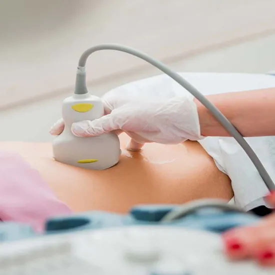
Looking for a reliable whole abdomen ultrasound near Vasant Kunj? At our diagnostic centre, we offer high-precision abdominal ultrasound scans performed by experienced radiologists using advanced equipment. This non-invasive imaging procedure is designed to assess vital organs and structures within your abdominal cavity, ensuring accurate and timely diagnosis. During the scan, a clear gel is applied to your abdomen to allow smooth transmission of high-frequency sound waves through a handheld device called a transducer. These sound waves create real-time images of your internal organs, which are displayed on a monitor for analysis.
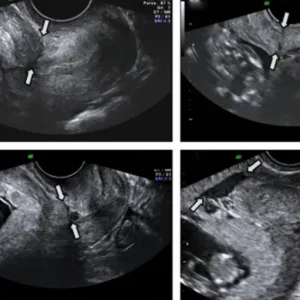
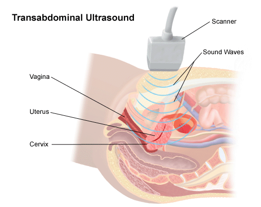
Ultrasound TVS (Transvaginal Scan)
Ultrasound Transvaginal Scan (TVS) is a specialized imaging technique primarily used in gynecology to examine the pelvic organs, particularly in women. During a TVS, a small transducer probe is inserted into the vagina, allowing for close proximity to the reproductive organs such as the uterus, ovaries, and fallopian tubes. This proximity enables high-resolution imaging, providing detailed views of the pelvic anatomy. TVS is commonly employed to diagnose conditions such as ovarian cysts, uterine fibroids, endometrial abnormalities, and to monitor early pregnancies. It is often preferred for its ability to produce clearer images compared to abdominal ultrasound, especially in cases where precise visualization is crucial. While the procedure may cause minor discomfort, it is generally safe, minimally invasive, and offers valuable diagnostic information for various gynecological conditions.
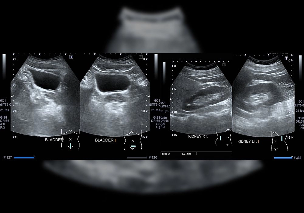
Ultrasound KUB (Kidneys, Ureters, Bladder)
Ultrasound Kidneys, Ureters, Bladder (KUB) is a non-invasive imaging modality focused on assessing the urinary system within the abdomen. Utilizing high-frequency sound waves, a KUB ultrasound examines the kidneys, ureters, and bladder, aiding in the diagnosis of a variety of urinary tract disorders. During the procedure, a handheld transducer is moved across the abdominal surface, capturing detailed images of the organs and their surrounding structures. KUB ultrasound is commonly used to detect conditions such as kidney stones, urinary tract infections, congenital abnormalities, and tumors. It offers the advantage of real-time imaging without the need for ionizing radiation, making it a safe and effective diagnostic tool for evaluating urinary tract health.
Ultrasound Lower Abdomen/Pelvis
Ultrasound Lower Abdomen/Pelvis is a diagnostic procedure used to examine the pelvic organs and structures situated in the lower abdominal region. By employing high-frequency sound waves, this non-invasive imaging technique provides detailed visualizations of organs such as the uterus, ovaries, fallopian tubes, bladder, and prostate gland in men. During the examination, a transducer is gently maneuvered over the skin surface of the lower abdomen, producing real-time images displayed on a monitor. Ultrasound of the lower abdomen/pelvis is commonly employed to evaluate conditions such as pelvic pain, abnormal bleeding, ovarian cysts, uterine fibroids, and prostate enlargement. It offers the advantage of being safe, painless, and does not expose patients to ionizing radiation, making it an essential tool in diagnosing a wide range of pelvic and abdominal disorders.
Ultrasound Upper Abdomen
Ultrasound Upper Abdomen is a diagnostic imaging technique utilized to examine the organs and structures located in the upper abdominal region. By emitting high-frequency sound waves, this non-invasive procedure creates detailed images of organs such as the liver, gallbladder, pancreas, spleen, and kidneys. During the examination, a transducer is placed on the skin surface of the upper abdomen, emitting sound waves that bounce off the internal organs, creating real-time images displayed on a monitor. Ultrasound of the upper abdomen is commonly employed to diagnose conditions such as liver disease, gallstones, pancreatitis, spleen abnormalities, and kidney disorders. It offers the advantage of being safe, cost-effective, and does not expose patients to ionizing radiation, making it a valuable tool in the evaluation of various upper abdominal pathologies.
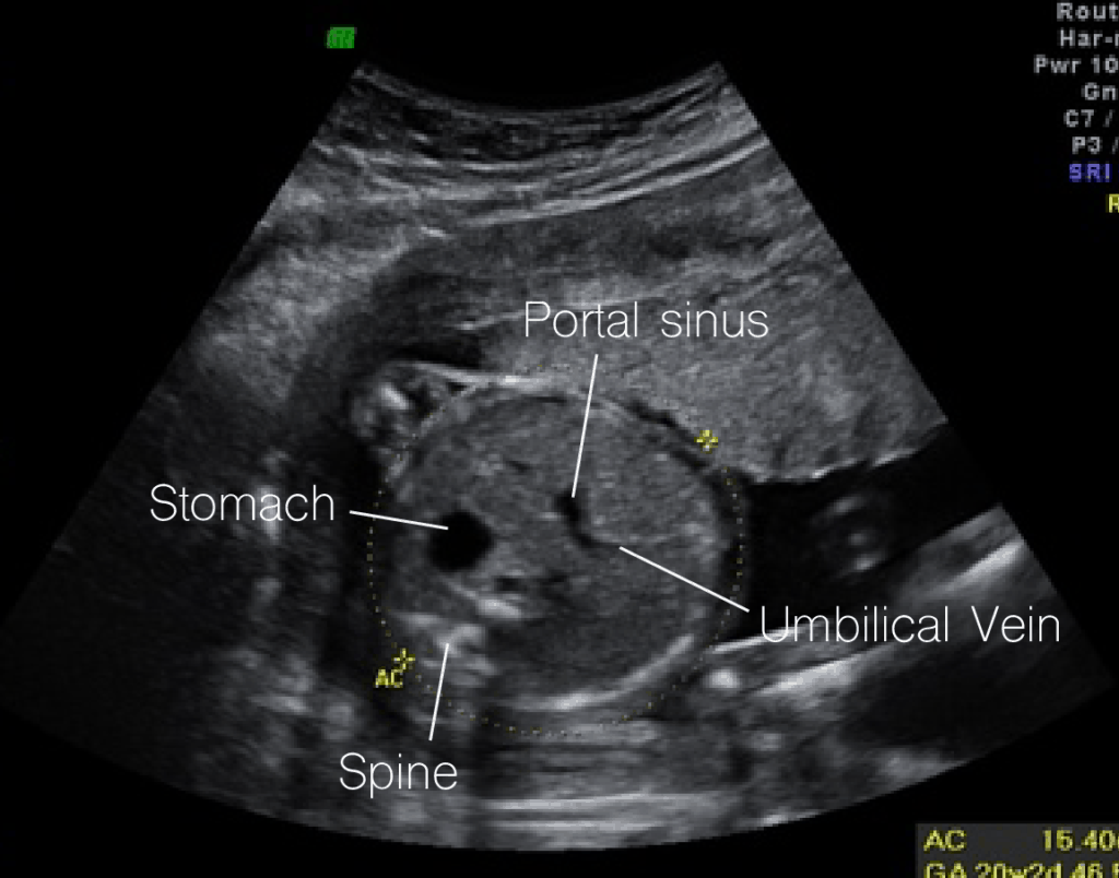
Ultrasound Level 2/TIFA Scan
Ultrasound Level 2/TIFA (Targeted Imaging for Fetal Anomalies) Scan is a comprehensive fetal ultrasound examination typically performed between 18 to 22 weeks of pregnancy. This advanced imaging technique aims to assess the detailed anatomy and development of the fetus, focusing on detecting any potential abnormalities or congenital defects. During the scan, a trained sonographer carefully examines various fetal structures, including the brain, spine, heart, limbs, and internal organs, to ensure they are developing normally. The Level 2/TIFA scan is particularly beneficial for identifying structural anomalies that may require further evaluation or intervention, providing crucial information for expectant parents and healthcare providers to make informed decisions about the management of the pregnancy.
Ultrasound Level 1/NT Scan
Ultrasound Level 1/NT (Nuchal Translucency) Scan is a specialized prenatal screening test typically conducted between 11 to 14 weeks of pregnancy. It involves measuring the thickness of the nuchal fold, a small pocket of fluid at the back of the fetus’s neck, to assess the risk of chromosomal abnormalities such as Down syndrome and other genetic conditions. Additionally, the scan evaluates other fetal markers, including the nasal bone and blood flow in the ductus venosus and tricuspid valve. By combining these measurements with maternal age and other factors, the Level 1/NT scan provides an estimate of the fetus’s risk for certain chromosomal abnormalities, aiding expectant parents and healthcare providers in making informed decisions about further diagnostic testing or pregnancy management.
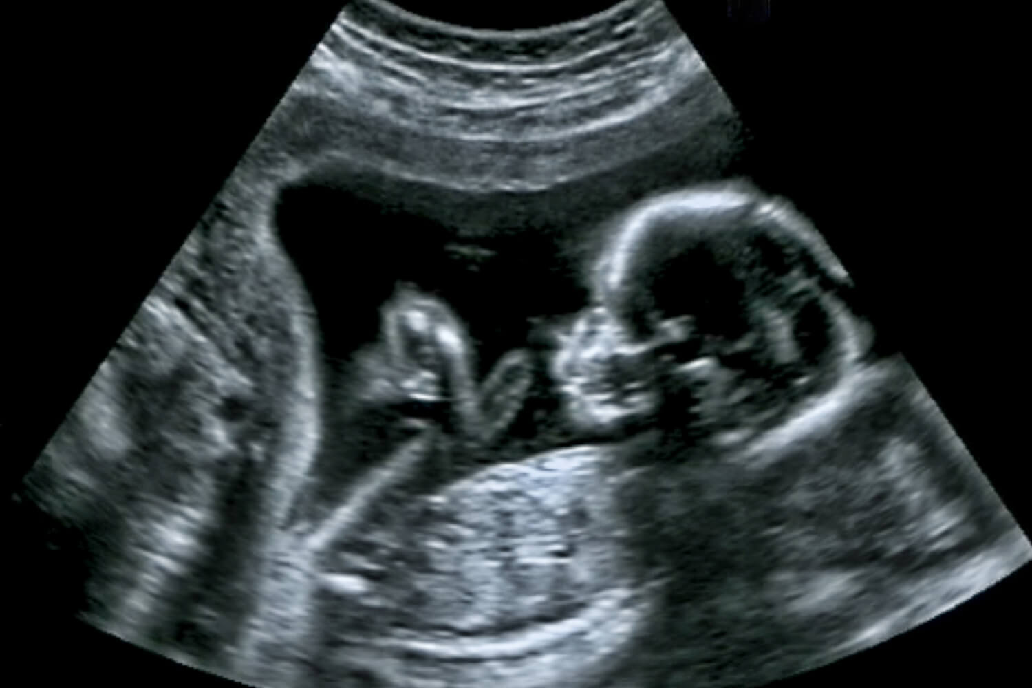
Ultrasound Obstetric Scan
Ultrasound Obstetric Scan is a pivotal diagnostic tool used throughout pregnancy to monitor the growth and development of the fetus, as well as to assess the health of the mother. This non-invasive imaging technique utilizes high-frequency sound waves to create real-time images of the fetus, placenta, and surrounding structures in the uterus. Obstetric ultrasound scans are performed at various stages of pregnancy to determine gestational age, confirm fetal viability, assess fetal anatomy, monitor fetal growth and position, and detect any abnormalities or complications such as placenta previa, fetal malformations, or intrauterine growth restriction. These scans also play a crucial role in guiding prenatal care and management decisions, allowing healthcare providers to provide optimal care for both the mother and the developing fetus.
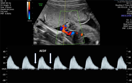
Color Doppler Obstetric Scan
Color Doppler Obstetric Scan is an advanced ultrasound technique used during pregnancy to assess the blood flow within the placenta, umbilical cord, and fetal blood vessels. By combining traditional ultrasound imaging with Doppler technology, which measures the speed and direction of blood flow, this scan provides valuable information about the vascular health of the placenta and fetus. It can help detect conditions such as placental insufficiency, fetal growth restriction, or abnormalities in umbilical cord blood flow, which may impact fetal well-being and pregnancy outcomes. Additionally, Color Doppler Obstetric Scan is utilized to monitor conditions like preeclampsia and to guide management decisions, ensuring optimal care for both the mother and the developing fetus throughout pregnancy.
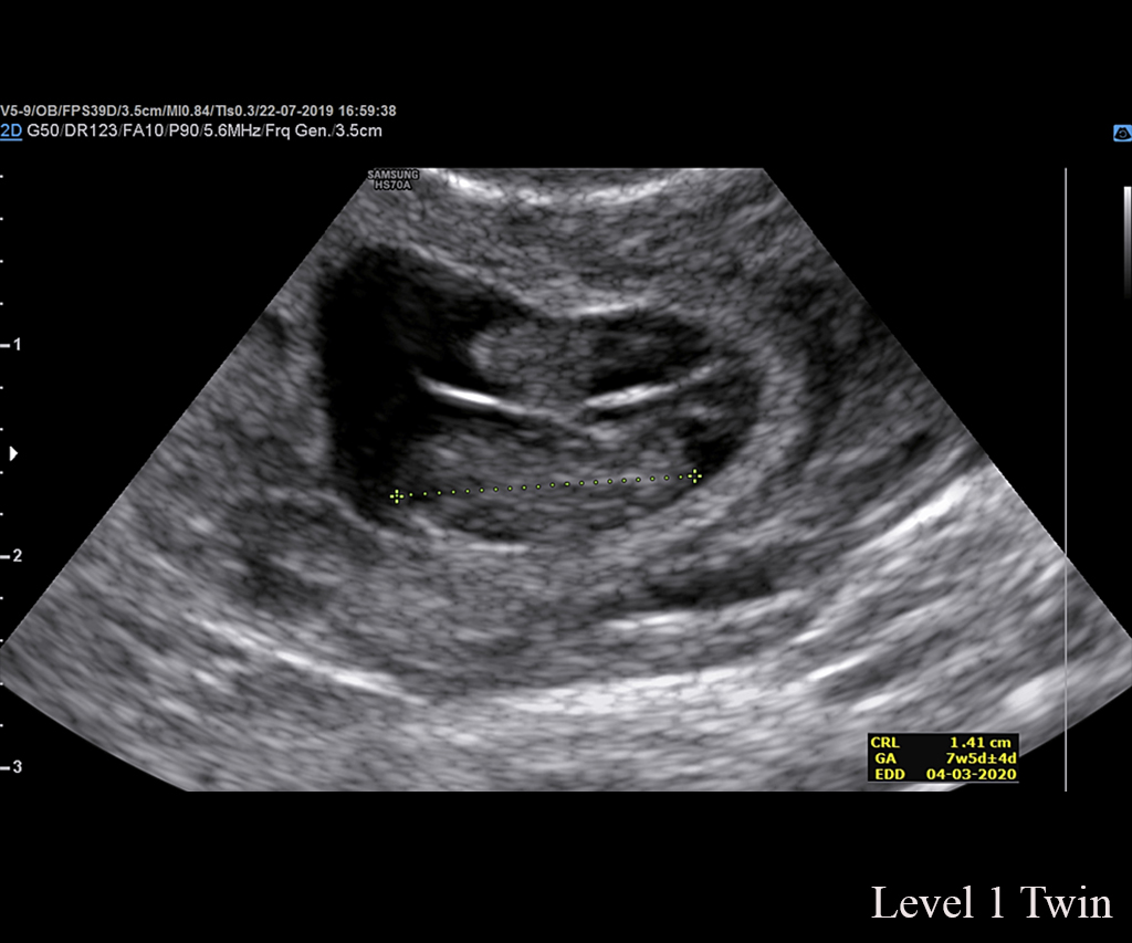
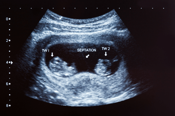
Ultrasound Level 2 (Twin Pregnancy)
Ultrasound Level 2 (Twin Pregnancy) is a specialized imaging procedure tailored for pregnancies involving twins or multiples. Conducted typically between 18 to 22 weeks of gestation, this comprehensive scan provides detailed assessments of the fetal anatomy, growth, and development of each individual fetus. With twins, it’s particularly important to evaluate factors such as the placental location, amniotic fluid levels, and any signs of twin-to-twin transfusion syndrome (TTTS) or other complications unique to multiple pregnancies. The Level 2 ultrasound for twin pregnancies helps healthcare providers identify any structural abnormalities, assess the chorionicity (whether the twins share a placenta), and guide management decisions to ensure the optimal health and well-being of both fetuses and the expectant mother throughout the pregnancy.
Follicular Monitoring Scans (Complete Study)
Follicular Monitoring Scans (Complete Study) involve a series of ultrasound examinations performed throughout the menstrual cycle to monitor the development and maturation of ovarian follicles. This specialized imaging technique is commonly used in assisted reproductive technologies (ART) and fertility treatments to optimize timing for procedures such as intrauterine insemination (IUI) or in vitro fertilization (IVF). During each scan, the size and number of developing follicles are assessed, along with changes in the endometrial lining. By tracking follicular growth and hormone levels, healthcare providers can determine the most fertile window for conception, adjust medication dosages if necessary, and maximize the chances of successful pregnancy. Follicular Monitoring Scans (Complete Study) play a crucial role in personalized fertility care, providing valuable insights into ovarian function and guiding treatment strategies to help couples achieve their reproductive goals.
Follicular Monitoring Scan (Single Study)
A Follicular Monitoring Scan (Single Study) is a specialized ultrasound examination conducted during a specific phase of the menstrual cycle to assess the development and maturation of ovarian follicles. This procedure is commonly utilized in fertility treatments to monitor follicular growth and predict ovulation timing accurately. During the scan, the size and number of developing follicles are evaluated, providing valuable information about the ovarian response to fertility medications and the potential for successful ovulation and conception. By performing a single study, healthcare providers can determine the optimal timing for interventions such as timed intercourse or insemination, maximizing the chances of achieving pregnancy while minimizing the need for multiple scans and healthcare visits. Follicular Monitoring Scan (Single Study) offers a streamlined approach to fertility monitoring, providing crucial information to support individualized treatment plans and enhance reproductive outcomes for couples undergoing fertility treatment.

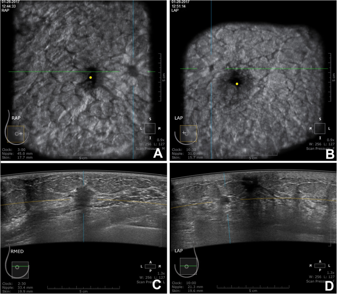
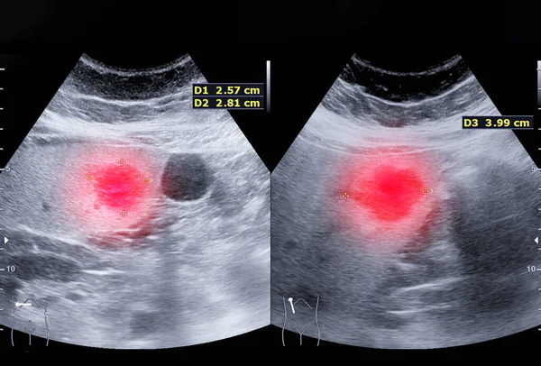
USG Both Breast Scan
Ultrasound (USG) Both Breast Scan is a specialized imaging examination used to evaluate both breasts for any abnormalities or changes in breast tissue. This non-invasive procedure employs high-frequency sound waves to create detailed images of the breast tissue, including the mammary glands, ducts, and surrounding structures. By examining both breasts simultaneously, radiologists can compare any differences in breast tissue and identify any suspicious findings such as masses, cysts, or areas of increased vascularity. USG Both Breast Scan is commonly used as a supplementary imaging tool alongside mammography or as a primary screening method for women with dense breast tissue or those at higher risk for breast cancer. It plays a crucial role in early detection, diagnosis, and monitoring of breast conditions, ultimately contributing to better outcomes and management of breast health.
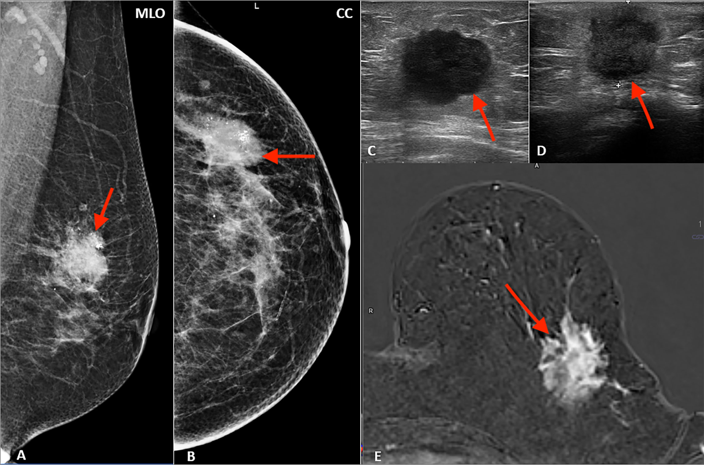
USG Single Breast Scan
Ultrasound (USG) Single Breast Scan is a focused imaging procedure used to examine a specific breast for any abnormalities or changes in breast tissue. This non-invasive technique utilizes high-frequency sound waves to produce detailed images of the breast, including the mammary glands, ducts, and surrounding structures. By concentrating solely on one breast, radiologists can closely evaluate any identified masses, cysts, or areas of concern, aiding in the diagnosis and management of breast conditions such as tumors, cysts, or fibroadenomas. USG Single Breast Scan is often recommended when a specific area of interest is identified during a clinical breast examination or mammography, providing additional information to guide further evaluation or treatment decisions.

USG Neck
Ultrasound (USG) Neck is a specialized imaging examination used to assess the structures within the neck region, including the thyroid gland, lymph nodes, salivary glands, blood vessels, and muscles. This non-invasive procedure employs high-frequency sound waves to create detailed images of the neck anatomy, aiding in the diagnosis of various conditions such as thyroid nodules, enlarged lymph nodes, cysts, abscesses, or vascular abnormalities. USG Neck is particularly valuable for evaluating thyroid disorders, guiding fine-needle aspiration biopsies, monitoring nodular growth, and assessing the extent of lymph node involvement in cases of infection or malignancy. With its ability to provide real-time imaging and precise localization of abnormalities, USG Neck plays a crucial role in the diagnosis and management of neck pathologies, ultimately facilitating timely and targeted treatment interventions.
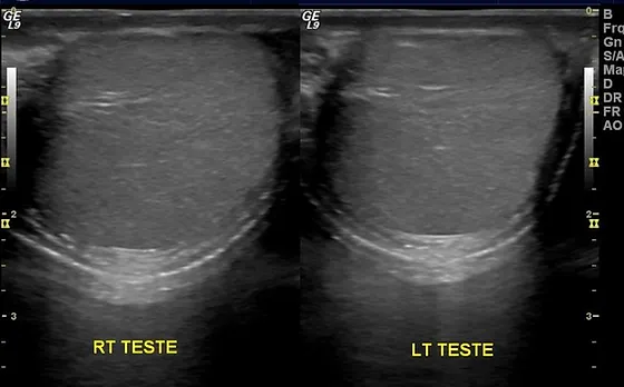
USG Scrotum
Ultrasound (USG) Scrotum is a diagnostic imaging procedure specifically designed to evaluate the structures within the scrotum, including the testicles, epididymis, and surrounding tissues. This non-invasive technique utilizes high-frequency sound waves to generate detailed images of the scrotal contents, aiding in the diagnosis of various conditions such as testicular torsion, epididymitis, hydrocele, varicocele, testicular masses, or other abnormalities. USG Scrotum is particularly valuable for assessing scrotal pain, swelling, or suspected testicular pathology, providing essential information for accurate diagnosis and appropriate management decisions. With its ability to visualize the internal structures of the scrotum with high resolution and clarity, USG Scrotum is an essential tool in the evaluation and monitoring of scrotal disorders, ensuring optimal care and outcomes for patients.
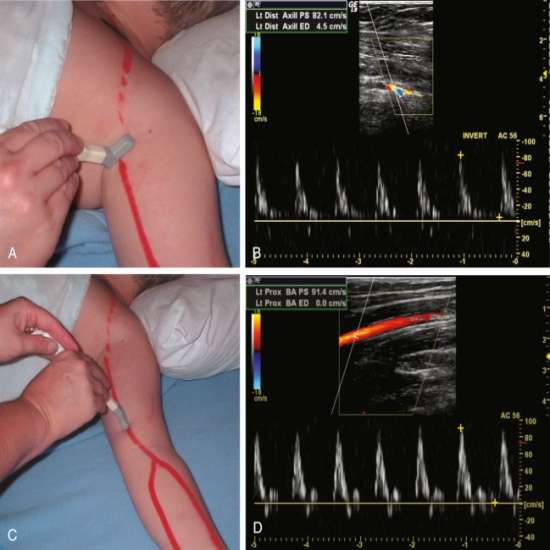
Bilateral Lower/Upper Arterial/Venous Colour Doppler
Bilateral Lower/Upper Arterial/Venous Colour Doppler is a specialized ultrasound technique used to evaluate the blood flow within the arteries and veins of the lower or upper extremities. This non-invasive imaging procedure combines traditional ultrasound imaging with Doppler technology to visualize and assess the direction, speed, and characteristics of blood flow in the peripheral vessels. By examining both sides of the body simultaneously, healthcare providers can compare blood flow patterns and detect abnormalities such as arterial stenosis, venous insufficiency, thrombosis, or peripheral artery disease. Bilateral Lower/Upper Arterial/Venous Colour Doppler plays a crucial role in diagnosing vascular disorders, guiding treatment decisions, and monitoring the effectiveness of interventions such as angioplasty, stenting, or vascular surgery, ultimately improving patient outcomes and vascular health.
Single Lower/Upper Arterial/Venous Colour Doppler
Single Lower/Upper Arterial/Venous Colour Doppler is a diagnostic ultrasound technique used to assess the blood flow within the arteries or veins of a specific limb or area of interest. This non-invasive imaging procedure combines conventional ultrasound imaging with Doppler technology to visualize and evaluate the direction, velocity, and characteristics of blood flow in the targeted blood vessels. By focusing on a single limb or area, healthcare providers can accurately diagnose conditions such as peripheral artery disease, deep vein thrombosis, or arterial or venous insufficiency. Single Lower/Upper Arterial/Venous Colour Doppler aids in the precise localization of vascular abnormalities, guiding treatment decisions, and monitoring the response to interventions such as medication, compression therapy, or surgical procedures, ultimately improving patient outcomes and vascular health.